IMCA Insights – October 2010
Visit at the UoA Electron Microprobe
Laboratory
by Anne Black
Quite a while ago
Dolores Hill (Senior Research Specialist, Lunar and Planetary Sciences,
University of Arizona, Tucson, Arizona) asked me if she could borrow
some thin-sections of Almahata Sitta from me for a very important
occasion. Of course, she could! In fact I had asked my favorite
thin-section maker to double-polish but leave some thin-sections
uncovered so they could be used for research. And I had just received
the first batch.
Then about a month ago, Dolores finally told me what that big occasion
was: the University had finally received their brand new, cutting-edge,
Electron Microprobe. It had taken a long time, but it was there, and had
been installed and tested by a couple of French technicians
(incidentally, I learned that the best Electron Microprobes are made in
France). The Lunar and Planetary Lab wanted to celebrate the event, and
have an official inauguration and ribbon-cutting ceremony; and I was
invited. And my thin-sections would be used for the "First Beam" because
of Almahata Sitta's connection with Tucson.
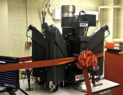
The big day was Friday September 24. When I arrived, the Lab was already
crowded, I met there Dr. Michael J. Drake, Director of the Lunar and
Planetary Laboratory, Dr. Kenneth Domanik, Manager of the Electron
Microprobe Laboratory, Dr. Dante Lauretta, Dolores Hill, and of course
Rik Hill, Richard Kowalski (who discovered Almahata Sitta when it was
still a meteor, and known as 2008 TC3), and many others. The huge
machine was ready and decorated with a large red bow; the big cylinder
in the middle is the electron gun, the smaller tubes coming out at
angles are the individual spectrometers, and the bow actually covers the
airlock where the sample is inserted.
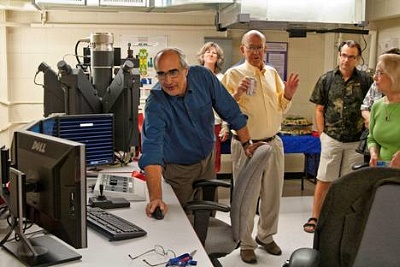
Dr. Domanik and Dr.
Drake promptly cut the ribbon and
Dr. Domanik settled down to show us what it could do
Photo courtesy of Full Moon Photography
Left to right: Dr.
Domanik, Maria Haas, Dr. Drake,
Dr. Dante Lauretta, Anne Black
Photo courtesy of Full Moon Photography
In case you don't know how a microprobe works, I will quote from the information kindly provided by Dolores Hill: "In an electron microprobe, a solid sample (on a thin-section) is bombarded with a focused beam of high energy electrons. This produces a variety of different types of interactions between the beam of electrons and the atoms in the sample. The result is a chemical analysis of the selected spots on the sample". For a more technical and complete explanation, please go to the following PDF files, kindly provided by Dolores Hill:
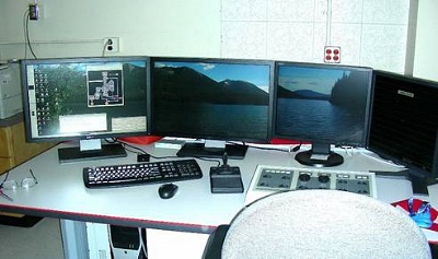
Photo by Anne Black
From a lay-person's point of view, there are 4 screens beside the microprobe proper, the user types in the various commands on a keyboard, and navigates around the thin-section with a joy-stick and a few knobs on another board. And Dr. Domanik took us on a tour of a thin-section of EL6 Almahata Sitta. First we saw a relict chondrule, black and white, and polarized.
EL6 Almahata Sitta
Chondrule in black and white
Photo by Dr. Domanik
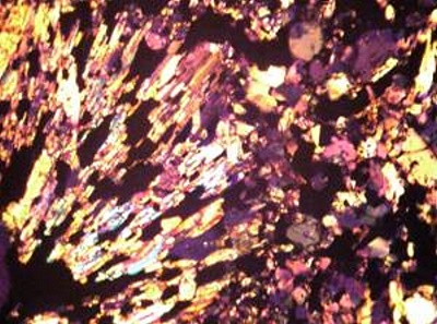
Photo by Dr. Domanik
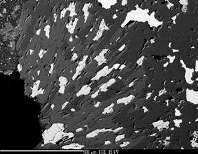
Then on to a tiny sliver of the crust. Dr. Domanik asked for the composition of a tiny light grey dot, and the machine promptly responded on another screen with the whole composition broken down in colorful peaks and valleys. What was it? Olivine. Then to two other intriguing stick-like, very dark masses, again the response came up quickly: Graphite.
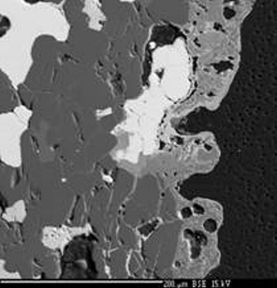
Photo by Dr. Domanik
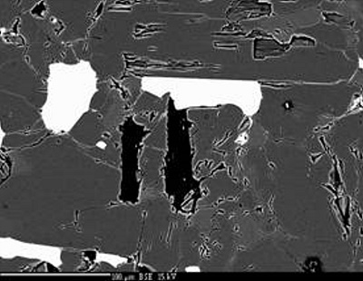
Photo by Dr. Domanik
For a more detailed,
and more scientific, explanation, provided by Dolores Hill, go to:
AlmahataSittaEL6.pdf
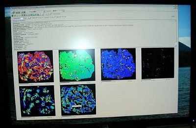
Then we went on to a
thin-section of one of the ureilite lithology of Almahata Sitta, and Dr.
Domanik demonstrated another function of the microprobe, X-ray mapping.
He asked for the distribution of several chemical elements, and the
machine promptly responded with colorful maps, each one showing the
location of one of the elements being searched. Again more detailed and
scientific information is available here:
AlmahataSittaURE.pdf
He then repeated the search on a thin-section of the Los Angeles
meteorite. Not only do the "maps", pictured below show the presence or
absence of the element being searched, but variations in the color also
indicate how prevalent that mineral is, a little to a lot. A very
precise and thorough analysis, however that does take longer for the
machine to produce, and this was an example Dr. Domanik had prepared.
More detailed information here:
LosAngeles.pdf
A thin section of the Los
Angeles shergottite under the microprobe
Photo by Anne Black
The Open House and
Inauguration was officially from 4 to 7 pm but we were all listening and
watching until way past 7; in fact few of us even took the time to
nibble on the finger-food kindly provided by the University. Sorry
Dolores, but some of us, and mostly me don't get very often the occasion
to look at an electron microprobe close-up and personal.
If you want to know more about the Lunar and Planetary Laboratory of the
University of Arizona, please look at:
AboutTheLab.pdf
As for me, I am delighted I came. Thank you very much to the whole
Laboratory team for the kind invitation.
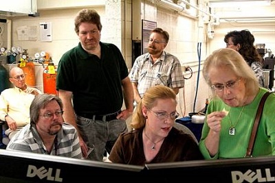
Left to right: Dr.
Drake, Richard Kowalski, Alex Gibbs,
Rik Hill,
Dolores Hill, Anne Black, Amy Phillips
Photo courtesy of Full Moon Photography
•
IMCA Home Page •
IMCA Code of Ethics •
IMCA Member List
•
Join IMCA •
IMCA Meteorite Info